Preoperative tumor size measurement in breast cancer patients: which threshold is appropriate on computer-aided detection for breast MRI?, Cancer Imaging
Background Computer-aided detection (CAD) can detect breast lesions by using an enhancement threshold. Threshold means the percentage of increased signal intensity in post-contrast imaging compared to precontrast imaging. If the pixel value of the enhanced tumor increases above the set threshold, CAD provides the size of the tumor, which is calculated differently depending on the set threshold. Therefore, CAD requires the accurate setting of thresholds. We aimed to compare the diagnostic accuracy of tumor size measurement using MRI and CAD with 3 most commonly used thresholds and to identify which threshold is appropriate on CAD in breast cancer patients. Methods A total of 130 patients with breast cancers (80 invasive cancers and 50 ductal carcinoma in situ [DCIS]) who underwent preoperative MRI with CAD and surgical treatment were included. Tumor size was manually measured on first contrast-enhanced MRI and acquired by CAD using 3 different thresholds (30, 50, and 100%) for each tumor. Tumor size measurements using MRI and CAD were compared with pathological sizes using Spearman correlation analysis. For comparison of size discrepancy between imaging and pathology, concordance was defined as estimation of size by imaging within 5 mm of the pathological size. Concordance rates were compared using Chi-square test. Results For both invasive cancers and DCIS, correlation coefficient rho (r) between tumor size on imaging and pathology was highest at CAD with 30% threshold, followed by MRI, CAD with 50% threshold, and CAD with 100% threshold (all p < 0.05). For invasive cancers, the concordance rate of 72.5% at CAD with 30% threshold showed no difference with that of 62.5% at MRI (p = 0.213). For DCIS, the concordance rate of 30.0% at CAD with 30% threshold showed no difference with that of 36.0% at MRI (p = 0.699). Compared to MRI, higher risk of underestimation was noted when using CAD with 50% or 100% threshold for invasive cancers and when using CAD with 100% threshold for DCIS. Conclusion For CAD analysis, 30% threshold is the most appropriate threshold whose accuracy is comparable to manual measurement on MRI for tumor size measurement. However, clinicians should be aware of the higher risk of underestimation when using CAD with 50% threshold for tumor staging in invasive cancers.

KJR :: Korean Journal of Radiology

year-old woman with true-positive breast MRI finding. Contrast-enhanced
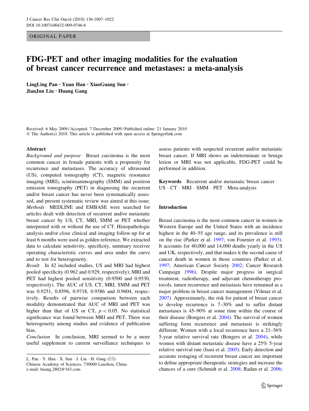
FDG-PET and other imaging modalities for the evaluation of breast
Higher underestimation of tumour size post-neoadjuvant chemotherapy with breast magnetic resonance imaging (MRI)—A concordance comparison cohort analysis
Preoperative tumor size measurement in breast cancer patients: which threshold is appropriate on computer-aided detection for breast MRI?, Cancer Imaging

Differentiating Grade in Breast Invasive Ductal Carcinoma Using Texture Analysis of MRI

Lipid-weighted intraoperative photoacoustic tomography of breast tumors: Volumetric comparison to preoperative MRI - ScienceDirect

The accuracy of magnetic resonance imaging in predicting the size of pure ductal carcinoma in situ: a systematic review and meta-analysis

Breast Magnetic Resonance Imaging for Patients With Newly Diagnosed Breast Cancer: A Review

Enhancement of breast cancer on pre-treatment dynamic contrast-enhanced MRI using computer-aided detection is associated with response to neo-adjuvant chemotherapy - ScienceDirect

Differentiating Grade in Breast Invasive Ductal Carcinoma Using
Breast lesion detection through MammoWave device: Empirical detection capability assessment of microwave images' parameters