A) Preoperative intraoral periapical (IOPA) radiograph of 36. B) Post operative (IOPA) radiograph of 36. C) 1 month follow up IOPA radiograph of 36. D) 6 months follow up IOPA radiograph of

A) Preoperative intraoral periapical (IOPA) radiograph of 36. B) Post operative (IOPA) radiograph of 36. C) 1 month follow up IOPA radiograph of 36. D) 6 months follow up IOPA radiograph of 36. E) 1 year follow up IOPA radiograph of 36. - IP Indian J Conserv Endod - clinical and preclinical conservative /restorative de

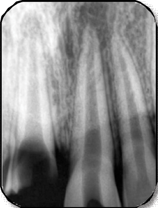
IOPA radiograph of mandibular molar region

Association between concentration of active MMP‐9 in pulpal blood and pulpotomy outcome in permanent mature teeth with irreversible pulpitis – a preliminary study - Sharma - 2021 - International Endodontic Journal - Wiley Online Library

JaypeeDigital

PDF) Direct pulp capping with bioactive materials – A case series
Management guidelines for amelogenesis imperfecta: a case report and review of the literature, Journal of Medical Case Reports

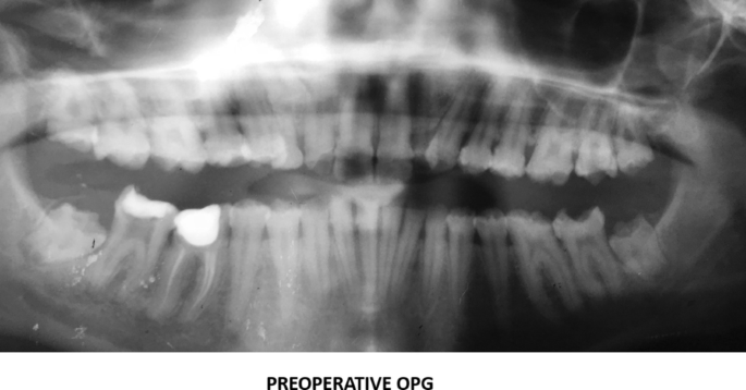
Role of Platelet-rich Plasma in the Healing of Impacted Third Molar
Coatings, Free Full-Text

a) The IOPA radiograph shows preoperative condition of the patient

A) Preoperative radiograph of tooth #46. (B) Postopera
jcdr-11-ZD05-g006.jpg