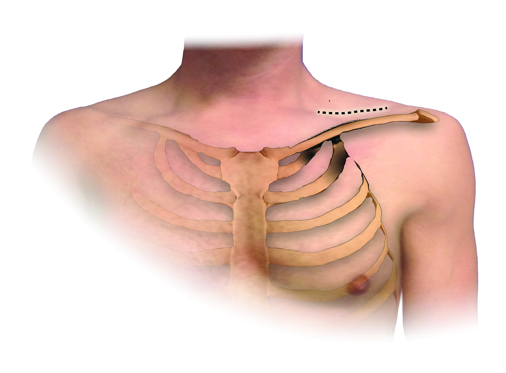
Figure 3 from Descriptive anatomy of the interscalene triangle and

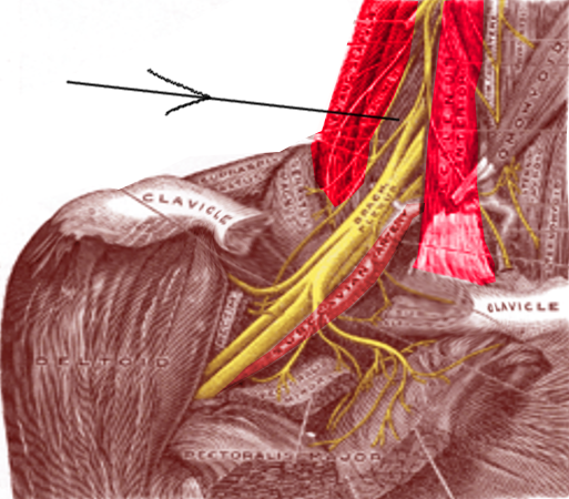
Fig 3. Depiction of the costoclavicular space. The neurovascular elements of the costoclavicular space can be seen here traveling superior to the first rib and inferior to the clavicle. The arrow indicates where measurements were taken. - "Descriptive anatomy of the interscalene triangle and the costoclavicular space and their relationship to thoracic outlet syndrome: a study of 60 cadavers."

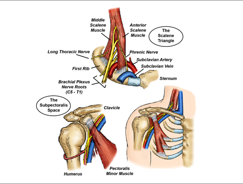
Anatomy of brachial plexus

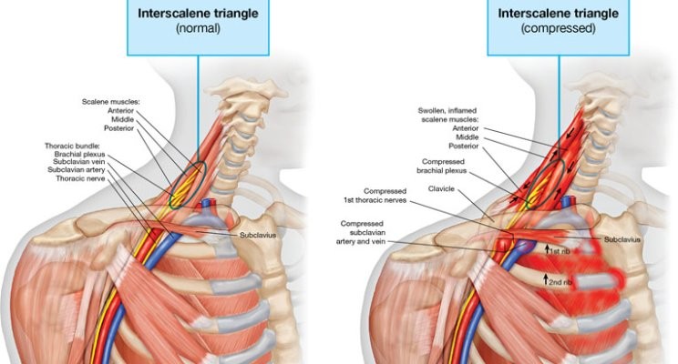
Neurogenic Thoracic Outlet Syndrome - ScienceDirect

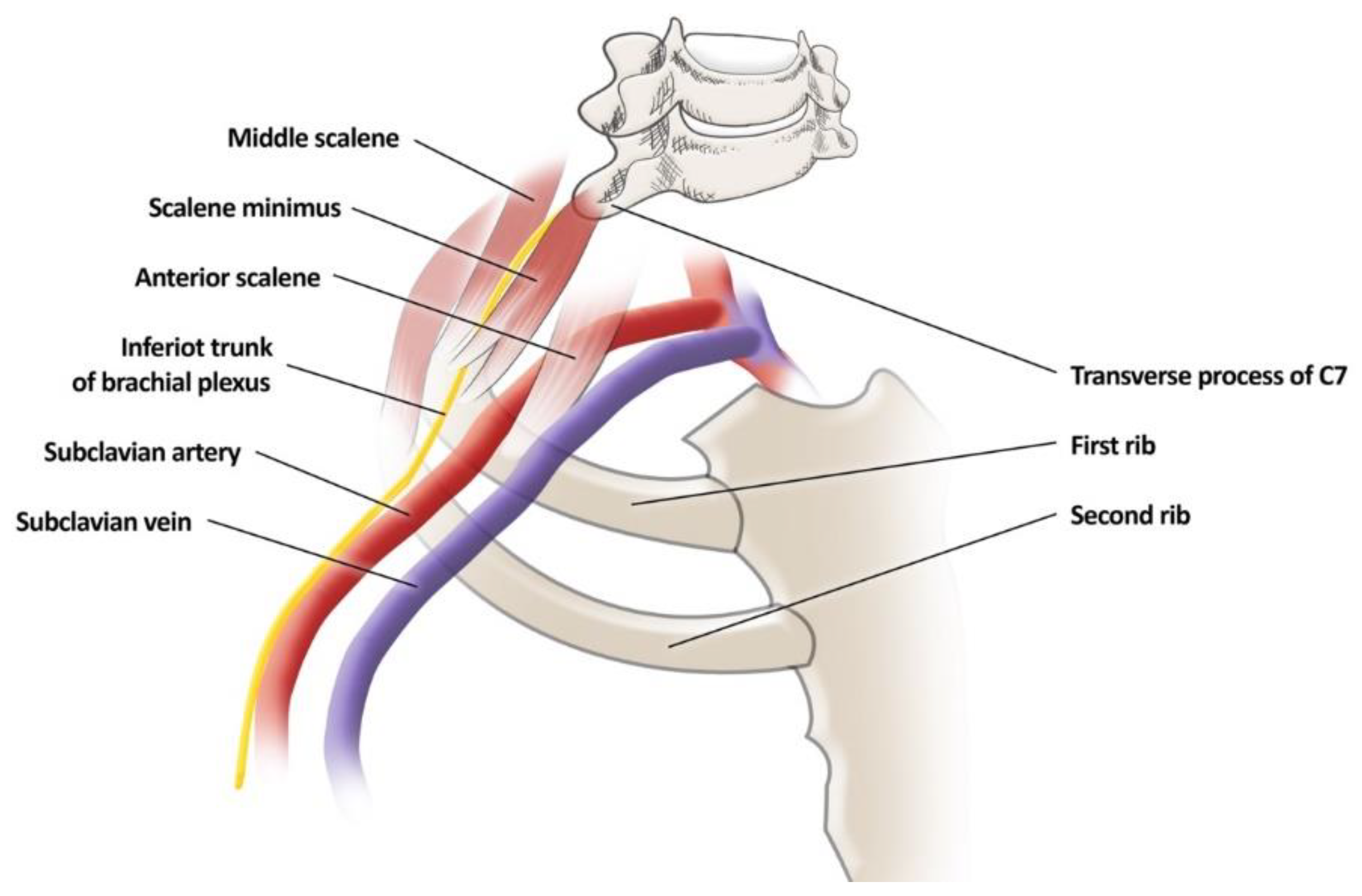
Overview of the structures of the thoracic outlet. There several

Imaging of non-specific complaints of the arm, neck, and/or shoulder (CANS): role of the scalene muscles and piercing variants in neurogenic thoracic outlet syndrome - ScienceDirect
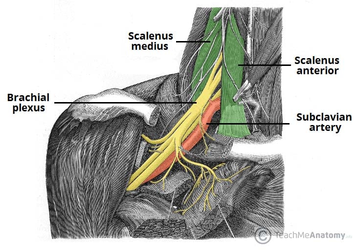
The Brachial Plexus - Sections - Branches - TeachMeAnatomy

Schematic diagram of surface anatomical measurements. a=length of

Surgical Neurology International
JCM, Free Full-Text

Reporting standards of the Society for Vascular Surgery for thoracic outlet syndrome - ScienceDirect

CB-724 Triangles of the Head and Neck Flashcards
Anatomy, Head and Neck: Inter-scalene Triangle, Treatment & Management

Descriptive Anatomy of the Interscalene Triangle and the Costoclavicular Space and Their Relationship to Thoracic Outlet Syndrome: A Study of 60 Cadavers - ScienceDirect

Daniel Clearfield, DO, MS, FAOASM on LinkedIn: Hydrodissection for the Treatment of Vascular Thoracic Outlet Syndrome

Thoracic Outlet Syndrome